What is Maculopapular Rash?
Maculopapular rash is a compound word composed of the words “macule” and “papule.”
A macule is a flat blemish or discoloration that measures less than 1 cm. A papule is as elevated lesion measuring less than 1 cm. Combining the two terms, a maculopapular rash is as a smooth skin rash or redness covered by elevated bumps [1, 2, 3].
Other names for it are morbilliform eruption or exanthematous eruption (exanthema).
Due to the presence of inflammation, maculopapular rash appears as reddened or erythematous. The distribution may be concentrated on a certain area of the body or it can be generalized. It may be acute, subacute, or chronic. Acute maculopapular rash lasts less than 4 weeks; 4-8 weeks for subacute; and it is considered chronic if the rash lasts for more than 8 weeks.
The occurrence of maculopapular rash for children is generally due to viral infection. For the adults, drugs and allergies are often the culprit.
Maculopapular rash is associated with a lot of factors. In the absence of fever, maculopapular rash is not necessarily a problem. But if the patient is febrile and has some other signs and symptoms, maculopapular rash might be a sign of a serious disease so do not ignore it [4].
Pictures of Maculopapular Rash
Photo Source: pediatricsconsultant360.com
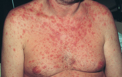
Picture 2: Maculopapular rash on HIV patient
Photo Source: primehealthchannel.com
Picture 3: Maculopapular rash in a patient with H1N1 influenza.
Image Source: cmaj.ca
Picture 4: A maculopapular rash seen in a man with severe diarrhea
Photo Source: archderm.jamanetwork.com
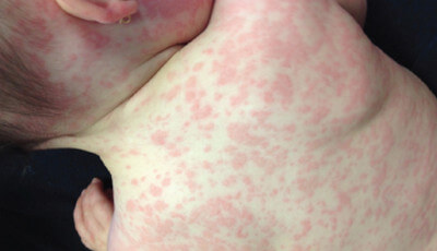
Picture 5: Maculopapular rash after otitis media
Image Source: pediatricsconsultant360.com
Picture 6: Erythematous Maculopapular Rash
Image Source: globalskinatlas.com
Picture 7: Maculopapular Rash image
Photo Source: path.upmc.edu
Picture 8: Maculopapular Rash on trunk
Photo Source: bestpractice.bmj.com
Classification by Etiology
Causes of Maculopapular Rash: Classification
Infectious Causes
Rubeola (Measles)
Maculopapular rash due to measles is common among children. It begins appearing on the hairline and behind the ears then it spreads through your trunk and extremities. When the rash desquamates and becomes brownish, it means it is starting to fade away. The order on which it appeared follows the order on when it fades away. The Koplik’s spots disappear after the onset of rash.
Rubella (German Measles)
Maculopapular rash caused by Rubella starts to appear on the face then it spreads downwards.
Roseola (Exanthema Subitum)
This occurs in children less than 3 years old. The maculopapular rash in this case is scattered although the face is often unaffected. It appears after the fever disappears.
Chikungunya Virus (Dengue)
In dengue, the maculopapular rash appears after the redness and itchiness of the skin. It spreads from the trunk to the face and limbs. Petechiae may also be noted.
Parvovirus B19 (Erythema Infectiosum or Fifth Disease)
Children who are 3-12 years old are commonly infected by this disease. Maculopapular rash in erythema infectiosum appears bright red that makes them look like they were slapped on the face. This is due to the fever that occurs before the appearance of the rash.
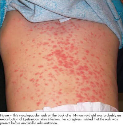
Infectious Mononucleosis
Older children and adolescents are affected by this kind of infection. The morbilliform rash can be seen on the trunks and arms [5].
Others
- Rickettsia rickettssi (Rocky Mountain Spotted Fever)
- Cytomegalovirus (CMV)
- Epstein-Barr Virus (EBV)
- Enteroviruses
- Treponema pallidum (Secondary Syphilis)
- Borrelia burgdorferi (Lyme Disease)
- Rickettsia prowazekii (Louse-Borne Typhus)
- Rickettsia typhi (Murine Typhus)
- Salmonella typhi (Typhoid Fever)
- Chlamydia psittaci (Psittacosis)
- Streptobacillus moniliformis (Rate-Bite Fever)
- Spirillum minus (Rate-Bite Fever)
- Leptospira (Leptospirosis)
- Ehrlichia and Anaplasma (Ehrlichiosis) [6]
Drug Eruptions
Medications such as antibiotics, anticonvulsants, and allopurinol may include maculopapular rash as one of its adverse reactions. There was a research on 50,000 patients and 91% of the drug eruptions presented itself as maculopapular eruption. Noted risk factors are immunosuppression, old age, and female gender.
Maculopapular rash is a type IV or delayed cell-mediated reaction. It is due to the body’s hypersensitivity of a drug or its metabolite.
The maculopapular rash is suspected to be due to drugs when it appeared within 4-12 days on the start of administering the new medication. It may be confirmed when you discontinue the usage of the drug and the maculopapular rash disappears within a certain amount of time. It reappears once the drug that you have an adverse reaction to is being readministered.
Anaphylaxis
Maculopapular rash may appear due to allergies to food and insect bite or sting. It only takes a while when the eruption starts.
Systemic and Rheumatologic Diseases
Acute graft-versus-host disease (stem cell or bone marrow transplant), Kawasaki disease (mucocutaneous lymph node syndrome), and Still’s disease (juvenile rheumatoid arthritis) present maculopapular rash as one of their signs and symptoms [4].
Maculopapular Rash in Children
In children, the main cause of maculopapular rash is viral exanthems such as rubeola, rubella, parvovirus B19, roseola, Epstein-Barr virus (EBV), cytomegalovirus (CMV), and adenoviruses [7].
Maculopapular Rash in Patients with HIV
In patients who are HIV positive, maculopapular rash presents as one of the early signs of the virus. It is a form of primary HIV infection that occurs in any parts of the body especially the face, trunk, and palms of the hand. In the mouth and genitals, maculopapular rash poses as ulcers. It lasts for 2-3 weeks [8, 9].
Diagnostic Approach
For the physicians, this diagram may be helpful for you to determine the cause of maculopapular rash. It guides you on how to proceed and what to do next in order to give the patient appropriate treatment.
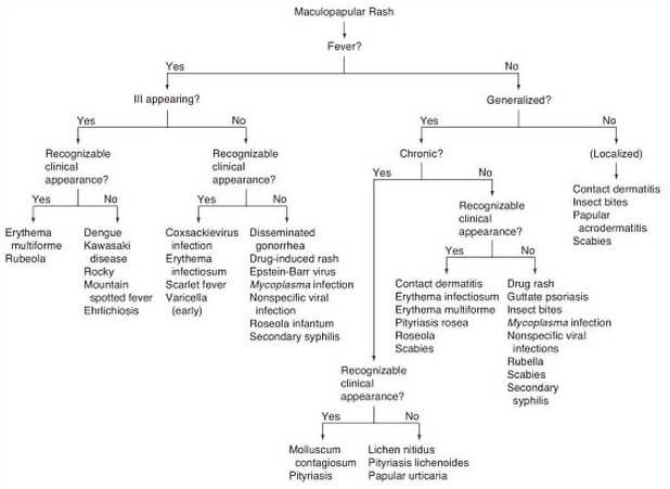
Picture 11 : Diagnostic Approach to Maculopapular Rash
Photo Source: Fleisher GR & Ludwig S, Textbook of Pediatric Emergency Medicine, Lippincott Williams & Wilkins 2010, p 511
In the presence of maculopapular rash, the first question to answer is if the patient has a fever. If yes, determine if they appear ill or not. If they do, the patient may either have erythema multiforme, rubeola, dengue, Kawasaki disease, rocky mountain spotted fever or Ehrlichiosis.
If they do not appear ill, what they have may be Coxsackie virus, erythema infectiosum, scarlet fever, varicella, gonorrhoea, drug-induced rash, Epstein-Barr virus, Mycoplasma infection, Roseola infantum, secondary syphilis, or viral infection.
If the patient is afebrile, determine if the rash is generalized or localized. If localized, the maculopapular rash may be due to contact dermatitis, insect bite, scabies, or papular acrodermatitis.
If generalized, determine the chronicity. If chronic, the patient may have molluscum contagiosum, pityriasis, lichen nitidus, pityriasis lichenoides, or papular urticaria. If acute, the patient may have contact dermatitis, erythema infectiosum, erythema multiforme, pityriasis rosea, roseola, scabies, drug rash, guttate psoriasis, insect bite, Mycoplasma infection, rubella, scabies, secondary syphilis, or other viral infections.
Differential Diagnosis of Maculopapular Rash
Maculopapular rash may be mistakenly called macule, papule, patch, nodule, plaque, vesicle, or pustule. To differentiate these skin lesions from each other, here are the pictures of each.
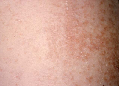
 Picture 12: Macule measures less than 1 cm. It is a flat blemish like that of a freckle.
Photo from Wright State University Dermatology
Image Source: antimicrobe.org
Picture 13: A patch is like an enlarged macule. It measures more than 1 cm.
Photo from Wright State University Dermatology
Photo Source: antimicrobe.org
Photo from Wright State University Dermatology
Photo Source: antimicrobe.org
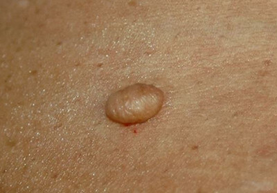
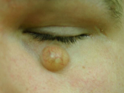
Picture 15: A nodule is a spherical elevation of the skin measuring more than 1 cm. It becomes a tumor once the size exceeds 2-3 cm.
Photo from Wright State University Dermatology
Photo Source: antimicrobe.org
Picture 16: A plaque measures more than 1.5 cm. The center area is flat and the edges have elevated lesions. An example of a plaque is psoriasis.
Photo from Wright State University Dermatology
Photo Source: antimicrobe.org
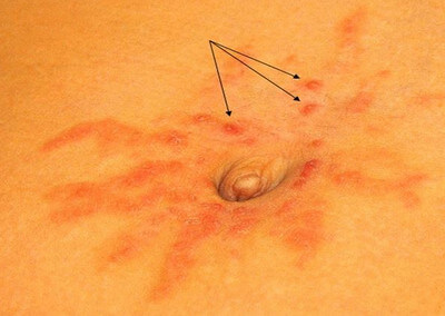
Picture 17: Vesicles (green arrows) and bulla (red arrow) both contain fluid in them. They are rounded and elevated. The difference between the two is the size. Vesicles measure less than 1 cm while bullae are more than 1 cm.
Photo from Wright State University Dermatology
Photo Source: antimicrobe.org
Picture 18: A pustule is a lesion that contains pus.
Photo from Wright State University Dermatology
Photo Source: antimicrobe.org
Click : Here is one awesome “mnemonic” about the Maculopapular rash
Maculopapular Rash Treatment
If maculopapular rash is caused by drugs, discontinue using it unless its benefits outweigh the risks. For patients who have penicillin allergy, avoid usage of carbapenems and cephalosporins. If the patient has allergies to a radioactive medium, use prednisone, diphenhydramine, and ephedrine or histamine H2-receptor antagonist before introducing it [8].
Maculopapular rash is not a medical condition by itself. Eliminate the cause in order to completely get rid of it.Treatment is large symptomatic. Antibiotics, antivirals, intravenous infusion, oral hydration, proper hygiene, and rest are necessary for patients who have infectious diseases.
Topical corticosteroids and oral antihistamines may relieve the discomfort [11]. Treatment may also include gamma and ultraviolet radiation. Patients with HIV and other advanced conditions need specialized care.
References
- Emory C, Maculopapular Rash, Junct 2011
- http://dermatology.about.com/cs/viralinfections/g/macpap.htm
- http://medical-dictionary.thefreedictionary.com/maculopapular+rash
- Naftanel M, Evaluation of Maculopapular Rash accessed on https://online.epocrates.com/u/2911774/Evaluation+of+maculopapular+rash/Differential/Overview
- Parthasarathy A, Textbook of Pediatric Infectious Diseases, JP Medical Ltd. 2013, p 68
- www.antimicrobe.org/e8.asp
- Identifying Infectious Disease in Children accessed on http://www.stacommunications.com/journals/diagnosis/2001/08_august/chawla.pdf
- Early Stage Symptoms of HIV accessed on https://www.dred.com/uk/early-symptoms-of-
hiv.html - Primary HIV Infection accessed on http://www.patient.co.uk/doctor/Primary-HIV-Infection.htm
- Riedl MA & Casillas AM, Adverse Drug Reactions: Types and Treatment Options, American Family Physician 2003 accessed on http://www.aafp.org/afp/2003/1101/p1781.html#sec-7
- Morbilliform Drug Eruption accessed on http://dermnetnz.org/reactions/morbilliform.html

