Definition of Ecchymosis
Ecchymosis [pronunciation: (/ˌɛkᵻˈmoʊsᵻs/, ICD-10: R23.3] is defined as hemorrhagic blotching due to pooling of blood under the skin or mucous membrane caused by medical conditions, hematologic diseases, or trauma.
Violaceous discoloration of the skin is observed which is usually negative for pain, induration, and firmness [1, 2].
After the trauma, red blood cells extravasate from the blood vessels into the underlying tissue. Macrophages phagocytose the red blood cells and eventually be degraded. The component of red blood cell that is responsible to violaceous color is hemoglobin, which will be enzymatically converted into bilirubin (blue green). Later on, bilirubin will be converted to hemosiderin which will give a golden brown color, when the ecchymosis is about to heal [3].
Ecchymosis Pictures
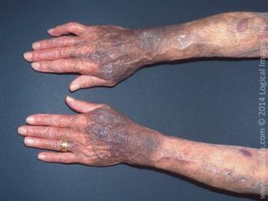
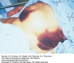
 Picture 1: Ecchymoses
Source: library.med.utah.edu
Picture 2: Ecchymosis in steroid-induced purpura
Source: VisualDx
Picture 3
Source: MDDK.com
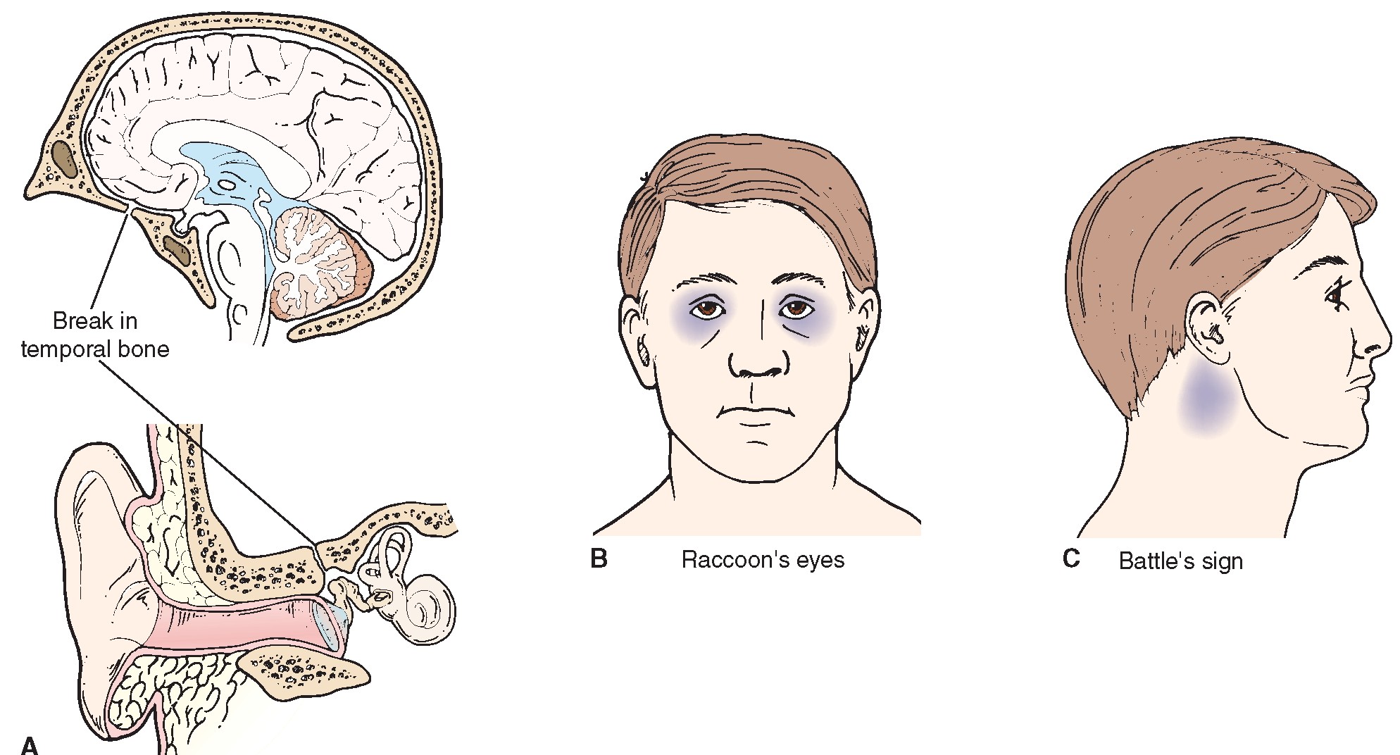
Picture 4: Basilar skull fracture in the temporal bone can cause raccoon’s eyes and Battle’s sign
Source: In Depth Tutorials and Information

Picture 5: Periorbital ecchymosis due to trauma
Source: MDDK.com
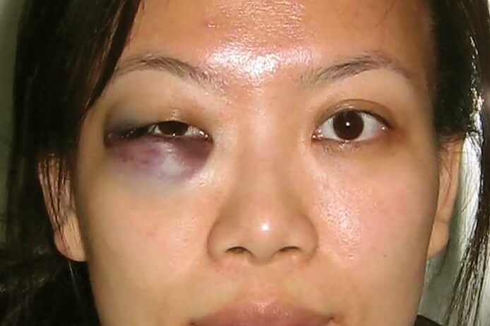
Picture 6: Unilateral periorbital ecchymosis
Source: Healthool
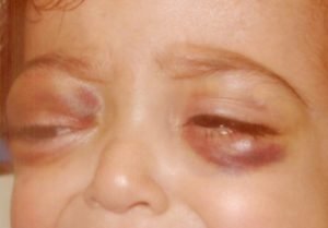
Picture 7: Bilateral periorbital ecchymosis
Source: MDDK.com
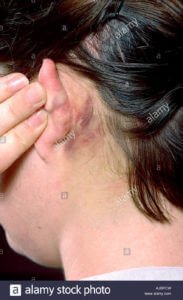
Picture 8: Battle sign or mastoid ecchymosis due to basilar skull fracture
Source: Alamy
Picture 9: Illustration of Cullen sign (periumbilical ecchymosis) and Grey Turner sign (ecchymosis on the lateral abdominal wall)
Source: Anesthesia Key

Picture 10: Petechiae vs. ecchymosis vs. hematoma
Source: StudyBlue
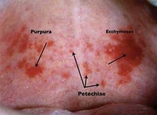
Picture 11: Purpura vs. petechiae vs. ecchymosis
Source: RDH Magazine
Signs Related to Ecchymosis
Raccoon Eye Sign or Blepharohematoma
Periorbital ecchymosis is most commonly associated with eyeball injury. It easily develops even with mild injury because of the loose connection between the skin of the eyelid and its underlying tissues [4,5].
Raccoon eye sign or blepharohematoma refers to periorbital ecchymosis which is usually due to trauma on the frontal area of the skull, leading to rupture of the veins of anterior cranial fossa. Blood then seeps into the periorbital area, hence the periorbital ecchymosis. Raccoon eye sign is a reliable sign for basilar skull fracture and/or intracranial lesions [6].
Compared to other parts of the body, ecchymosis around the eyes tends to be darker because of heavier pooling of the blood. Periorbital ecchymosis changes its color from purple to green to yellow until the blood underneath lyses. This may persist for around two weeks [4].
Battle Sign
Battle sign refers to retroauricular or mastoid ecchymosis primarily due to basilar skull fracture. Because of the trauma, the blood flow from emissary veins of the sigmoid sinus into the posterior auricular soft tissue is disrupted, hence the mastoid ecchymosis [6,7].
Cullen Sign
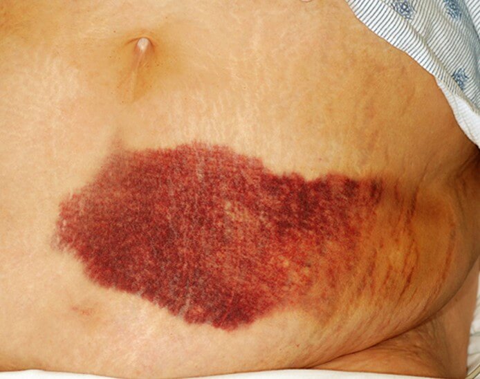
Cullen sign refers to periumbilical ecchymosis. Through the gastrohepatic and falciform ligaments, blood from the retroperitoneum collects around the umbilicus, giving it a violaceous color. The following are the causes of ecchymosis around the umbilicus [6]:
- Liver cirrhosis with portal hypertension
- Hemorrhaging ascites
- Percutaneous liver biopsy
- Hepatocellular carcinoma
- Strangulated umbilical hernia
- Acute pancreatitis
- Intrauterine pregnancy with bilateral acute salpingitis
- Ovarian cyst hemorrhage
- Splenic rupture
- Ruptured abdominal aortic aneurysm
- Hypothyroid myopathy
- Ileal strangulation with hemorrhage
- Ischemic and gangrenous bowel
- Perforated duodenal ulcer
- Retroperitoneal necrotizing fasciitis
- Renal sarcoma
Grey Turner Sign
Grey Turner sign refers to ecchymosis seen on the lateral side of the abdominal wall primarily among persons with acute pancreatitis. Other causes of Grey Turner sign are the following:
- Intrauterine pregnancy with bilateral acute salpingitis
- Sclerosing peritonitis
- Rectus sheath hematoma
- Retroperitoneal necrotizing fasciitis
- Ischemic and gangrenous bowel
- Ruptured abdominal aortic aneurysm
- Intra-aortic balloon pump insertion
- Cardiac catheterization
Through a defect in the transversalis fascia, extravasation of blood from the posterior pararenal space to the lateral side of quadratus lumborum occurs. The blood seeps through the abdominal wall and into the subcutaneous tissue, hence the ecchymosis on the flank. In the presence of Grey Turner sign, mortality rate is 40% [6].
Other Signs
- Crescent Sign: Unilateral or bilateral ecchymosis of the malleoli due to synovial rupture secondary to knee hemarthrosis.
- Stabler Sign: Ecchymosis on the inguinal-pubic area due to acute hemorrhagic pancreatitis, ruptured ectopic pregnancy, abdominal aortic aneurysm rupture, or neonatal adrenal hemorrhage.
- Fox Sign: Ecchymosis on the upper outer thigh due to acute suppurative pancreatitis, abdominal aortic aneurysm, strangulated ileum, pulmonary infarction, urethral instrumentation, or subcutaneous injection.
- Blue Scrotum Sign of Bryant: Ecchymosis in the scrotum due to abdominal aortic aneurysm. Blood passes through the inguinal canal and spermatic cord into the subcutaneous scrotal tissue.
- Seat Belt Sign: Ecchymosis with injury to the lumbar spine, abdominal organs, and/or thoracic organs associated with the use of seat belt after a motor vehicle accident. [6]
Causes of Ecchymosis
To diagnose the cause of ecchymosis, complete history taking and physical examination must be performed. In the presence of trauma, ecchymosis speaks for itself. But in the absence of it, a medical or hematologic condition is suspected. The person should then undergo coagulation panel test which includes prothrombin time, partial thromboplastin time, bleeding time, clotting time, platelet count, and thrombocyte aggregation assay [8].
Vitamin K Deficiency
One of the skin manifestations of vitamin K deficiency is ecchymosis, along with purpura and hemorrhage. This is due to decrease in vitamin K-dependent clotting factors II, VII, IX, and X. Treatment includes intramuscular injection of vitamin K 5-10mg/day for several days [9].
Snake Bite
Around 30 enzymes are found in the snake venom. It mostly comprises of hydrolases which causes the systemic and local effects of the snake bite. The site of the snake bite immediately develops skin redness, swelling, and ecchymosis. Antivenin and antitetanus would be of great help [9].
Bulimia
Self-induced vomiting is associated with bulimia. The dominant hand is put into the mouth until the gag reflex is stimulated and vomiting is induced. The hand becomes constantly rubbed against the teeth, resulting to Russell’s sign – crusted papules on the back of the hand. Clenching of the fist results to ecchymosis of the fingertips and bleeding underneath the nails [9].
Grain Itch
Grain itch is also known as barley itch, straw itch, prairie itch, or mattress itch. This is caused by mites seen on hay, barley, wheat, oat, and other cereals that is why farmers are mostly at risk. A small vesicle sits on top of an itchy reddish papule that is commonly seen on the trunk. Eventually, the lesion turns into an ecchymosis once the hemosiderin settles on the site [9].
End Stage Renal Disease
End stage renal disease with metastatic calcification results to calciphylaxis. This is described as a violaceous, mottled patch which eventually becomes a nodular, necrotic ecchymosis. This is usually seen on the thighs and abdomen. There were reports of gangrene and amputation due to its severity [9].
Treatment for Ecchymosis
In 2010, a study by Karen et al proved that a pulsed dye laser therapy is effective in healing ecchymosis [10]. But this is nothing but temporary. The ecchymosis may come back as long as the cause is not treated. Therefore, the cause of ecchymosis should be diagnosed to provide the correct treatment. However, supportive treatment like resting the affected area and putting ice on it may alleviate the pain or discomfort associated with the ecchymosis.
References
- Quinn CE. The Medical Record as a Forensic Resource. 2004.
- Gresele P, et al. Platelets in Hematologic and Cardiovascular Disorders: A Clinical Handbook. Cambridge University Press. 2007.
- Willy N. Ecchymosis. Culp Press. 2012.
- Fleisher GR & Ludwig S. Textbook of Pediatric Emergency Medicine 6th edition. Lippincott Williams & Wilkins. 2010.
- Mackenzie W. A Practical Treatise on the Diseases of the Eye. Longman, Brown, Green and Longmans. 1854.
- Epperla N, Mazza JJ, Yale SH. A Review of Clinical Signs Related to Ecchymosis. Wisconsin Medical Journal Volume 114 Number 2. April 2015.
- Caroline NL. American Academy of Orthopedic Surgeons. Nancy Caroline’s Emergency Care in the Streets, Volume 2. Jones & Bartlett Learning. 2007.
- Eversole LR. Clinical Outline of Oral Pathology: Diagnosis and Treatment 3rd edition. 2001.
- James WD, Berger TG, Elston DM. Andrews’ Clinical Diseases of the Skin Clinical Dermatology 10th edition. Elsevier, Inc. 2006.
- Karen JK, Hale EK, Geronemus RG. A Simple Solution to the Common Problem of Ecchymosis. Arch Dermatol. 2010;146(1):94-95. doi:10.1001/archdermatol.2009.343